Interpretation Summary
- Left ventricular size and systolic function are normal with an estimated EF of 60-65%.
- Right ventricular size and systolic function are normal.
- No echo evidence of valvular vegetation or associated significant regurgitation. Recommend TEE if clinical suspicion for endocarditis is high.
Study Comments:
A two-dimensional transthoracic echocardiogram with m-mode, color flow and Doppler was performed. The study was technically adequate with some images being suboptimal in quality. There is no prior echocardiogram noted for this patient. The patient was in a tachycardic rhythm during the exam.
Left Ventricle:
The left ventricle is grossly normal size. There is normal left ventricular wall thickness. Left ventricular size and systolic function are normal with an estimated EF of 60-65%. No regional wall motion abnormalities noted.
Right Ventricle:
The right ventricle is normal in size, thickness and function.
Left Atrium:
The left atrial size is normal.
Right Atrium:
The right atrium is normal in size.
Mitral Valve:
The mitral valve leaflets appear normal. There are no obvious valvular vegetations related to endocarditis seen on transthoracic echo. A transesophageal echo is suggested if clinically indicated. There is trace mitral regurgitation.
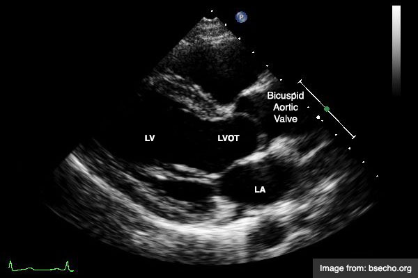
Aortic Valve:
The aortic valve is bicuspid but otherwise normal in structure and function. There are no obvious valvular vegetations seen. There is no aortic valve stenosis. There is trace aortic regurgitation.
Tricuspid Valve:
The tricuspid valve is grossly normal. There is trace tricuspid regurgitation.
Pulmonic Valve:
The pulmonic valve is not well seen, but is grossly normal. There is no pulmonic valvular regurgitation.
Great Vessels:
The aortic root is normal size. The inferior vena cava appeared normal and decreased < 50% with respiration (RAP 5-10mmHg).
Pericardium/Pleural:
There is no pericardial or pleural effusion.